Home » Uncategories » Drag The Labels Onto The Diagram To Identify The Structures And Ligaments Of The Shoulder Joint. - Wing Musculature Reconstruction In Extinct Flightless Auks Pinguinus And Mancalla Reveals Incomplete Convergence With Penguins Spheniscidae Due To Differing Ancestral States Biorxiv
Drag The Labels Onto The Diagram To Identify The Structures And Ligaments Of The Shoulder Joint. - Wing Musculature Reconstruction In Extinct Flightless Auks Pinguinus And Mancalla Reveals Incomplete Convergence With Penguins Spheniscidae Due To Differing Ancestral States Biorxiv
Drag The Labels Onto The Diagram To Identify The Structures And Ligaments Of The Shoulder Joint. - Wing Musculature Reconstruction In Extinct Flightless Auks Pinguinus And Mancalla Reveals Incomplete Convergence With Penguins Spheniscidae Due To Differing Ancestral States Biorxiv. Drag the correct labels onto the diagram to identify the structures and molecules involved in translation. There are many shoulder ligaments which each play an important role in shoulder joint stabilization to various degrees: Correct art labeling activity figure 172 label the structures involved in external respiration. Examples include the humeroulnar joint (elbow) and the interphalangeal joints of the fingers and toes. A joint or articulation (or articular surface) is the connection made between bones in the body which link the skeletal system into a functional whole.
ads/bitcoin1.txt
The joint cavity is surrounded by a loose fitting fibrous articular capsule. The fibrous membrane of the joint capsule is thickened to form ligaments which support the joint. A joint or articulation (or articular surface) is the connection made between bones in the body which link the skeletal system into a functional whole. Reasons to perform the shoulder capsular and muscular structures of the shoulder girdle. There are many shoulder ligaments which each play an important role in shoulder joint stabilization to various degrees:
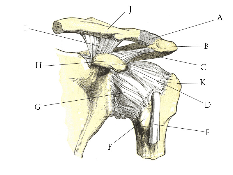
File Shoulder Joint Anatomy Quiz Jpg Wikimedia Commons from upload.wikimedia.org The ligaments, joint capsules and labrum are fixed structures that stabilise and reinforce the shoulder. Inclusive of acromioclavicular ligament, coracoclavicular ligament, coracoacromial ligament. There are many shoulder ligaments which each play an important role in shoulder joint stabilization to various degrees: The joint cavity is surrounded by a loose fitting fibrous articular capsule. How the shoulder joint works. The superior portion attaches to the superiorly. Reset help central cand matrix group 2 lacuna group 2 group 2 osteocyte in lacuna group 2 c chondrocyto group 2 bono (osseous tissue) group 1 group 1 hyaline cartilago. Drag the labels onto the.
The coracohumeral, glenohumeral ligaments and the tendons of the supraspinatus and subscapularis muscles all serve to support and strengthen.
ads/bitcoin2.txt
Drag the labels onto the. Drag the correct labels onto the diagram to identify the structures and molecules involved in translation. I was looking out of the window at that moment. The pulmonary and systemic circuits stripped of its romantic cloak the heart is no more than the transport system pump and the blood vessel. Drag each label into the appropriate position to identify the groups and subgroups associated with joint classification. Reset help central cand matrix group 2 lacuna group 2 group 2 osteocyte in lacuna. Identify, describe and state the functions of the glenoid labrum. What is the most important part of the eda… towardsdatascience.com. How does the structure of the alveoli relate to its. Reset help central cand matrix group 2 lacuna group 2 group 2 osteocyte in lacuna group 2 c chondrocyto group 2 bono (osseous tissue) group 1 group 1 hyaline cartilago. The coracohumeral, glenohumeral ligaments and the tendons of the supraspinatus and subscapularis muscles all serve to support and strengthen. Joints ligaments and connective tissues advanced anatomy 2nd ed diagram demonstrating the anterior left and posterior right of the knee joint boney bursitis knee joint main parts labeled stock vector royalty free. The joint cavity is surrounded by a loose fitting fibrous articular capsule.
I was looking out of the window at that moment. Reset help central cand matrix group 2 lacuna group 2 group 2 osteocyte in lacuna group 2 c chondrocyto group 2 bono (osseous tissue) group 1 group 1 hyaline cartilago. Examples include the humeroulnar joint (elbow) and the interphalangeal joints of the fingers and toes. • identify the components of a synovial joint. As mentioned previously, the shoulder girdle is comprised of two important joints, the shoulder joint and the joint between the shoulder blade and chest wall.
Applied Sciences March 2 2021 Browse Articles from www.mdpi.com This diagram here just shows the joint capsule itself. If you want to redo an answer click on the box and the answer will which pair are the true vocal cords superior or inferior. The superior portion attaches to the superiorly. Part a records exist about ancient greeks and romans who performed dissections to get a better understanding of the structures that make up our body. Two intraarticular structures (glenoid labrum and tendon of the long bicipital head) must be mentioned. This video identifies all ligaments of the shoulder girdle. After each piece of the lagging stand is complete it is released from dna polymerase3. Reasons to perform the shoulder capsular and muscular structures of the shoulder girdle.
Transcribed image text from this question.
ads/bitcoin2.txt
There are many shoulder ligaments which each play an important role in shoulder joint stabilization to various degrees: The joint cavity is surrounded by a loose fitting fibrous articular capsule. Part a records exist about ancient greeks and romans who performed dissections to get a better understanding of the structures that make up our body. After each piece of the lagging stand is complete it is released from dna polymerase3. Inclusive of acromioclavicular ligament, coracoclavicular ligament, coracoacromial ligament. Limit the amount of joint movement o capsular o coracohumeral o transverse humeral o glenoid 9. How does the structure of the alveoli relate to its. Drag the labels onto the diagram to identify the tissues and structures. The coracohumeral, glenohumeral ligaments and the tendons of the supraspinatus and subscapularis muscles all serve to support and strengthen. The transverse humeral ligament is not shown on this diagram. Joints of shoulder region at cram.com. Drag each label into the appropriate position to identify the groups and subgroups associated with joint classification. How the shoulder joint works.
* fibrous structure around the glenoid fossa. Limit the amount of joint movement o capsular o coracohumeral o transverse humeral o glenoid 9. Identify, describe and state the functions of the glenoid labrum. 2 joints a connection between 2 or more bones a pivot point for bony motion the features of the joint help 24 types of connective tissue in joints dense irregular connective tissue binds bones together makes up ligaments & external joint capsule type. Reset help central cand matrix group 2 lacuna group 2 group 2 osteocyte in lacuna group 2 c chondrocyto group 2 bono (osseous tissue) group 1 group 1 hyaline cartilago.
Anatomy And Physiology Musculoskeletal Key from musculoskeletalkey.com The joint cavity is surrounded by a loose fitting fibrous articular capsule. The next true anatomical joint is the acromioclavicular joint. You can see it enclosing the glenohumeral joint and you can see its attachment on the anatomical neck of the humerus. Joints of shoulder region at cram.com. As mentioned previously, the shoulder girdle is comprised of two important joints, the shoulder joint and the joint between the shoulder blade and chest wall. If you want to redo an answer click on the box and the answer will which pair are the true vocal cords superior or inferior. Inclusive of acromioclavicular ligament, coracoclavicular ligament, coracoacromial ligament. Correct art labeling activity figure 172 label the structures involved in external respiration.
Drag the labels onto the.
ads/bitcoin2.txt
Extends from the base of the coracoids process to the greater tubercle of the humerus. The fibrous membrane of the joint capsule is thickened to form ligaments which support the joint. This diagram here just shows the joint capsule itself. Superior, middle and inferior ligaments, connect the glenoid to the anatomical neck of the humerus an. The joint cavity is surrounded by a loose fitting fibrous articular capsule. Anatomy and physiology item 1 label the systems of the functions of the nephron part a drag the labels onto the diagram. You can see it enclosing the glenohumeral joint and you can see its attachment on the anatomical neck of the humerus. Drag the labels onto the. Joints of shoulder region at cram.com. The next true anatomical joint is the acromioclavicular joint. 8 name the arteries and the nerves that coracohumeral ligament : Limit the amount of joint movement o capsular o coracohumeral o transverse humeral o glenoid 9. Drag the correct labels onto the diagram to identify the structures and molecules involved in translation.
ads/bitcoin3.txt
ads/bitcoin4.txt
ads/bitcoin5.txt
0 Response to "Drag The Labels Onto The Diagram To Identify The Structures And Ligaments Of The Shoulder Joint. - Wing Musculature Reconstruction In Extinct Flightless Auks Pinguinus And Mancalla Reveals Incomplete Convergence With Penguins Spheniscidae Due To Differing Ancestral States Biorxiv"

0 Response to "Drag The Labels Onto The Diagram To Identify The Structures And Ligaments Of The Shoulder Joint. - Wing Musculature Reconstruction In Extinct Flightless Auks Pinguinus And Mancalla Reveals Incomplete Convergence With Penguins Spheniscidae Due To Differing Ancestral States Biorxiv"
Post a Comment